Orthopedic doctors specialize in the care of bones and the tissues that connect and operate them. This means the skeleton, ligaments, tendons, muscles, and nerves throughout the entire body. So, when an issue arises, diagnosis can be complicated and requires a skilled clinician such as those at AICA Lithia Springs. Diagnosis requires several steps, from a physical exam to imaging studies, all of which are offered at the office for your convenience.
Medical History
Medical history is one of the most critical parts of the orthopedic assessment. The story of how you injured yourself, called the history of the present illness (HPI), can be as relevant as the actual physical presentation of the injury. Tell the doctor when the pain started, how long it has lasted, and what kind of accident led to the injury. The kind of motions involved, if any, can often suggest what type of injury has occurred. For example, an accident that involves a twisting motion is more likely to result in an injury to the meniscus than a simple fall would be. Pain without a traumatic event is more likely to be related to an ongoing or acute disease process rather than an injury. The orthopedic doctor will also ask if there is anything you are doing at home that has brought you any relief or made the pain worse.
A detailed medical history that includes the patient’s prior medical diagnoses, surgeries, medications, activity level, nutritional status, and more can help the clinician narrow down the potential source of pain and identify contributing factors. Be sure to take a list of your current medications with you when you go to any orthopedic doctor’s appointment, including the name, the dose, the frequency you take the medication, and what route you take it by (oral, patch, etc.). Since some medications can weaken connective tissues, it is important to include any medications you have taken recently, even if you have stopped them, particularly antibiotics in the fluoroquinolone family or steroids. Always include any nutritional or herbal supplements you take, as well as over-the-counter medications such as acetaminophen.
Things to bring or do for your doctor’s appointment:
- A list of current and recent medications, supplements, and over-the-counter drugs.
- A list of all medical diagnoses, major injuries, and prior surgeries.
- Notes on what caused the injury or the course of the pain, anything that made it worse or better, and any home treatment being used.
- Wear loose and comfortable clothing with supportive shoes.
Physical Examination
It requires skill and experience to correctly assess a patient for orthopedic issues. The exam will usually begin with a visual observation of the body part that is affected, the unaffected part on the other side for comparison, and the rest of the body and how it relates to the injury. The orthopedic doctor may ask you to move the part in a particular way or walk on it to see if the movement or gait is normal. They will consider if the patient is able to put any weight on the extremity at all or if the limb is deformed, red, or swollen.
Range of motion is the extent to which any joint can bend out (abduction) and inward (adduction). During a physical examination, the orthopedic doctor may use a special kind of ruler to determine the exact range of motion your injured extremity has and compare it to the unharmed side. Changes in the range of motion can indicate an injury or deviation due to long-standing misuse or posture problems. For example, the neck can be easily injured in a car accident, reducing the range of motion, but looking down at your cell phone or tablet for hours each day can also cause a change in the range of motion as some muscles become weakened and others too tight.
It may be necessary for the orthopedic doctor to palpate the muscles and joints themselves, feeling for differences between sides and abnormal sounds such as grinding and popping. You may be asked to change position from lying down to sitting and then standing. Wear comfortable and supportive shoes so that you can walk if asked to.
There are specific maneuvers that providers are trained to perform that can elicit a response that indicates a certain type of injury is present. These maneuvers often require that the doctor touch your body and move the part themselves, so be sure to wear loose, comfortable clothing to the appointment. The doctor will ask permission before touching you, and you should be sure to let them know if anything is painful or if you need them to stop at any point. A good clinician will often have a solid idea about what problem they are facing after the history and exam are completed.
Diagnostic Testing
 While the history and physical exam are important, most orthopedic doctors will also order additional testing studies to confirm their suspicions. Imaging studies include radiographs (also called x-rays), computed tomography scans (CTs), and magnetic resonance imaging (MRI). The doctor may order lab testing to see if there are signs of certain diseases in the blood, like rheumatoid arthritis, which can affect joints.
When going for any testing:
- Wear loose and comfortable clothing. You may be asked to change into a gown for some tests.
- Take a list of all medications and supplements you use.
- Inform the technician if you are pregnant, diabetic, claustrophobic, and have any known drug or substance allergies.
- Read all instructions thoroughly before and after testing.
Radiographic Imaging
Radiography, also called X-ray, uses a small amount of radiation to obtain an image that is cast onto a special type of film. The denser a tissue, the whiter it appears on radiography, which means bones and heavy tissues are easier to see than delicate connective tissues. They are the most commonly used imaging tools for orthopedic issues and can range from simple to complex. The provider can take stationary pictures or moving images in a technique called fluoroscopy. They may choose to perform an arthrogram using fluoroscopy by injecting dye directly into the joint while observing the image in real-time.
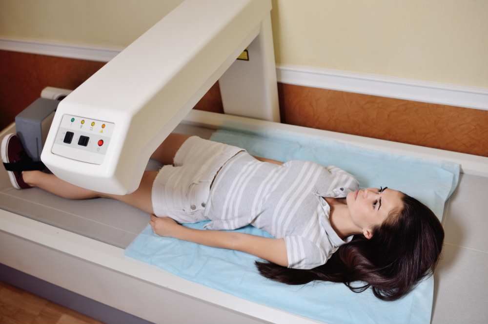
Bone Scan
Another form of radiographic imaging is the Dual-energy X-ray absorptiometry (DEXA) bone scan, which utilizes radioactive isotopes that bind to specific tissues that show up clearly when a special type of x-ray is done. This allows the clinician to see changes in the bone tissue itself, like tumors, infection, and thinning. The bone density scans older women have yearly to detect osteoporosis is an example of this kind of testing. The radioactive substance used is excreted readily from the body and is not harmful to the patient. These scans are particularly useful in diagnosing stress fractures which can be difficult to see on other forms of radiographs.
Ultrasonography
 Ultrasound has many benefits for assessing orthopedic issues since it is inexpensive, safe, and readily available. Using soundwaves that reflect off of tissues to create an image that can be seen in real-time, including with motion, the ultrasound offers the orthopedic doctor the ability to see both bones and soft tissues. It can be performed while the patient is moving, which is particularly useful for assessing how movement affects a joint. Ultrasound can also be used to check blood vessels for blood clots or atherosclerosis that can impair blood flow to tissues. The downside to ultrasound is that images are as clear as other forms of radiography, and it can be time-consuming.
Computed Tomography (CT)
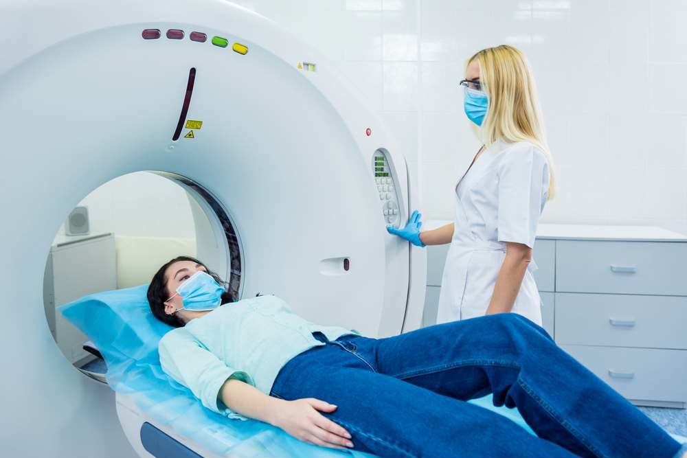 Like other radiographic testing, CT scans use a small amount of radiation; they can provide a two-dimensional image that includes soft tissues and blood vessels. Some advanced algorithms can actually produce a three-dimensional image of some scans as well. The orthopedic doctor may order a CT scan to be done with contrast, which can be given intravenously, orally, or injected directly into a joint (called an arthrogram). A dye can be injected into the disc of a spine in a test called a discogram. This can help diagnose diseases of the spinal discs that may require surgical repair. CT scans are relatively quick and painless, although some people find the intravenous line or dye injection mildly uncomfortable. Some people may also find the machine itself claustrophobic, but fortunately, there are options for open CT scanners that are less enclosed. Let the technician know if you are pregnant or allergic to iodine.
Magnetic Resonance Imaging (MRI)
MRI combines the best of ultrasonography and radiography while eliminating the negatives of both. MRI uses radiofrequency waves generated by a powerful magnet to create a crisp, clean image of all tissue types. Like the ultrasound, there is no radiation involved, but like the CT scan, it can give a three-dimensional image that shows amazing detail. As with CT, the orthopedic doctor may opt to perform this test using a dye injected into the joint or veins first. Specific radioisotopes can be used to tag certain tissues, similar to those used in the bone scan. The biggest downside is that MRIs can take a long time, and some people find the confined space of the machine itself to be uncomfortable. Some locations offer open MRIs that are less confining. Since radiofrequency waves are being used, the test also produces a lot of noise, and you will be asked to wear headphones and earplugs to protect your hearing.
Electromyogram (EMG)
The EMG is often performed when the orthopedic doctor suspects the issue is related to the nerves that control muscles. The test is generally done on a specific extremity, such as one arm, and is done using very fine needles that are inserted into the muscles of that part. Electrodes are attached to the needles that tell a machine how the nerves are functioning as you squeeze and relax each muscle. This test is often done in conjunction with nerve conduction studies, which use electrodes to measure how quickly electrical signals travel down the nerve. These tests are particularly useful in assessing for neuropathy and myopathy.
All testing for orthopedic issues should be done by a doctor who is skilled in the assessment and evaluation of the results.
The orthopedic doctors at AICA Lithia Springs have extensive experience in diagnosing and caring for patients with compassion. You can reach out to them today to make an appointment.